Radio From Scratch
Trusted education for medical learners

Video Lessons for Radiology Enthusiasts

Explore selected public preview videos. Full access to our complete radiology video library is reserved for verified members.

Chest radiograph
Preview
Members Library
5

public previews

Free to watch

Full catalog

for verified members

3+ lessons

Public Preview Videos

Watch these free preview lessons on the site.

Basics of Radiology (Medical imaging 101 series)-First Episode
Basics
Preview

Basics of Radiology (Medical imaging 101 series)-First Episode

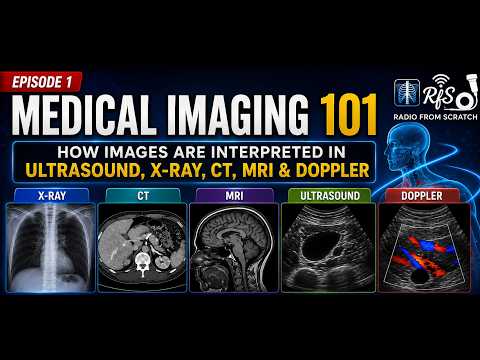
Welcome to the first episode of Basics of Radiology ( Medical Imaging 101 Series) ! In this episode , we will cover the fundamentals of radiology and introduce the basics principles of Medical Imaging. Stay tuned for future episodes as we explore each imaging modality in greater detail.
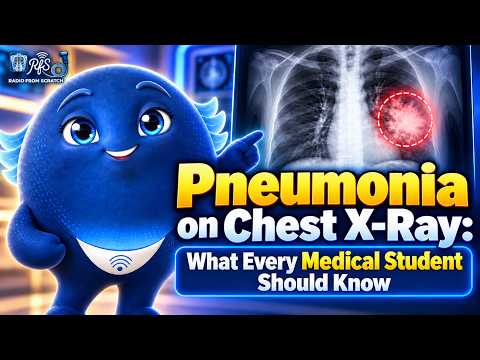
Pneumonia on Chest X-ray ( Part 1)
Cardiothoracic
Preview

Pneumonia on Chest X-ray ( Part 1)

Designed for medical students, IMGs, and anyone learning chest X-ray interpretation. Want to learn more? Sign up for a FREE membership to access exclusive educational videos and resources.

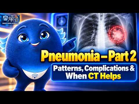
Pneumonia - Part 2
Cardiothoracic
Preview

Pneumonia - Part 2

Pneumonia on Chest X-Ray: Patterns, Complications & When CT Helps | Part 2 --Pneumonia doesn't always appear as classic lobar consolidation. It can be patchy, multifocal, complicated, or difficult to detect on the initial chest X-ray. --In this episode of Radio From Scratch, we explore different radiographic patterns of pneumonia, potential complications, and when CT imaging can help. -- What you'll learn: Recognizing patchy and multifocal airspace opacities . Identifying pleural effusion and cavitation . Understanding the limitations of chest X-rays . Knowing when CT imaging is helpful --- Designed for medical students, IMGs, and anyone learning chest X-ray interpretation. --Want to learn more? Sign up for a FREE membership to access exclusive educational videos.

Pneumothorax - Part 1
Cardiothoracic
Preview

Pneumothorax - Part 1

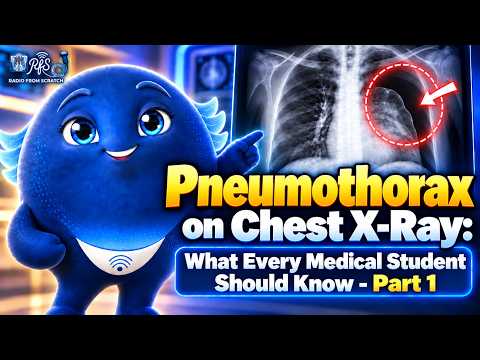
🫁 Pneumothorax on Chest X-Ray: What Every Medical Student Should Know | Part 1 -- A patient suddenly develops sharp chest pain and shortness of breath. Would you recognize a pneumothorax on the chest X-ray?--In this episode of Radio From Scratch, we explore the essential radiographic findings of pneumothorax and how to recognize them on chest X-rays.--What you'll learn: What a pneumothorax is and how it affects the lung.How to identify the visceral pleural line.Why lung markings disappear beyond the pleural line. How comparing both lungs helps identify pneumothorax-- Designed for medical students, IMGs, and anyone learning chest X-ray interpretation.--Want to learn more? Sign up for a FREE membership to access our exclusive educational videos. 📸 Image Credits & Attribution-- Image 1:Bonilla A, Blair AJ, Alamro SM, et al. “Recurrent spontaneous pneumothoraces and vaping in an 18-year-old man: a case report and review of the literature.” Journal of Medical Case Reports. 2019;13:283. Figure 1a. --Original image: https://link.springer.com/article/10.1186/s13256-019-2215-4/figures/1 --License: CC BY 4.0 — https://creativecommons.org/licenses/by/4.0/ --Modifications: Cropping, removal of original arrows, and reformatting for educational presentation.

Pneumothorax - Part 2
Cardiothoracic
Preview

Pneumothorax - Part 2

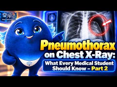
🫁 Pneumothorax on Chest X-Ray: What Every Medical Student Should Know | Part 2-- Pneumothorax isn't always easy to recognize on a chest X-ray, especially in patients lying supine. What subtle signs should you look for, and when does pneumothorax become a life-threatening emergency?-- In this episode of Radio From Scratch, we explore the radiographic findings of supine and tension pneumothorax and their clinical significance. 🔎 What you'll learn: - Why pneumothorax can be difficult to detect on supine chest X-rays - How to recognize the deep sulcus sign - Key imaging findings associated with tension pneumothorax - Why treatment of clinically apparent tension pneumothorax must not be delayed for imaging 🎓 Designed for medical students, IMGs, and anyone learning chest X-ray interpretation. 🌐 Want to learn more? sign up for a FREE membership to access our exclusive educational videos. These videos are unlisted on YouTube and can be accessed through our website. 📸 Image Credits & Attributions Image 1 – Right-Sided Pneumothorax--- Bonilla A, Blair AJ, Alamro SM, et al. “Recurrent spontaneous pneumothoraces and vaping in an 18-year-old man: a case report and review of the literature.” Journal of Medical Case Reports. 2019;13:283. Figure 1a.-- Original: https://link.springer.com/article/10.1186/s13256-019-2215-4/figures/1 --License: CC BY 4.0 — https://creativecommons.org/licenses/by/4.0/ --Modified by adding red arrows, cropping, and reformatting for educational presentation. --Image 2 – Deep Sulcus Sign --Hellerhoff. “Deep sulcus sign 59jm - Roe - 001.jpg.” Wikimedia Commons, 2018. --Original: https://commons.wikimedia.org/wiki/File:Deep_sulcus_sign_59jm_-_Roe_-_001.jpg --License: CC BY-SA 3.0 — https://creativecommons.org/licenses/by-sa/3.0/ --Modified by adding a red circle, arrow, text annotation, and reformatting. Adapted image shared under CC BY-SA 3.0. --Image 3 – Large Left Pneumothorax with Mediastinal Shift --Clinical Cases. “Pneumothorax CXR.jpg.” Wikimedia Commons, 2006. --Original: https://commons.wikimedia.org/wiki/File:Pneumothorax_CXR.jpg --License: CC BY-SA 2.5 — https://creativecommons.org/licenses/by-sa/2.5/ --Modified by adding red arrows, outlines, and annotations highlighting the pneumothorax, collapsed lung, and rightward mediastinal/tracheal displacement. Adapted image shared under CC BY-SA 2.5.

Why Sign Up?

Membership is for verified medical and medical-adjacent learners.

Full curated video catalog

Access the entire library of radiology lessons and cases across every subspecialty.

Watch after login

Member-only lessons play on the site after an access check. You can also open them on YouTube.